Mini C-Arm Machines: What Is It and How Do They Work
A mini C-arm can produce digital images instantly. It is a significant advantage compared to traditional X-ray imaging devices that use a film-based image intensifier and require a longer wait time for results. Ensure that the mini C-arm you’re considering is easy to use for your surgeons and radiographers. Ask for a demo or trial to see how it operates in a real-world setting.
Portable Fluoroscopic System
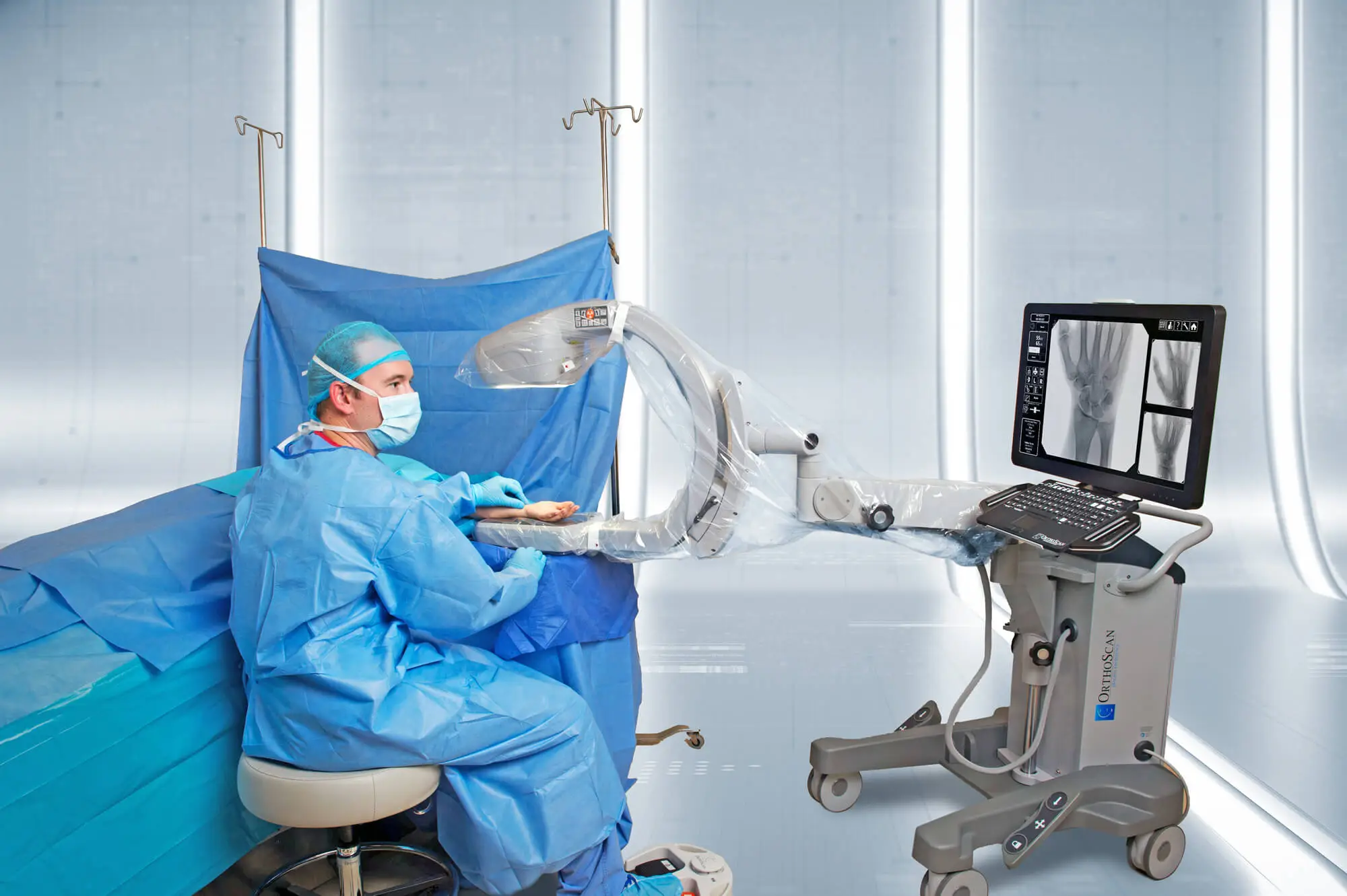
A mini C-arm is a portable fluoroscopic system that uses X-rays to create real-time images for orthopedic and surgical procedures. It’s ideal for hospital settings, pet clinics, pediatric orthopedics and sports medicine. Invented in 1955, this workhorse of medical imaging is named for its C-shaped arm that connects one end with an X-ray source and the other with a detector to capture pictures and record motion. The system is usually equipped with a flat-panel sensor, which delivers instant results and reduces radiation exposure compared to X-ray image intensifiers. It also features anti-scatter grids to help lessen the X-ray scatter effect during a procedure. When buying a mini C-arm from online vendors like minicarm.com, ask your surgeons and radiographers about the ease of use and user interface. Please find out how long it takes to complete the job and whether the machine requires additional training. In addition, ask how many push buttons it takes to get a specific result.
Non-Invasive Imaging Device
The smart-C mini C-arm x-ray machine is collapsible and portable, making it easily transported between rooms. It also offers flexibility for point-of-care imaging in the field. This device enables orthopedic surgeons to perform complex arthroscopic procedures on knees, hips, shoulders, and femur. Its rotating flat detector allows surgeons to capture images from multiple angles. It also provides a more precise viewing of long bones and reduces radiation exposure. The smallest footprint on the market enables this system to be moved easily between exam rooms, satellite clinics, urgent care centers, emergency departments, and athletic team venues. It can be used in the operating room and cath lab, making it a versatile tool for hospitals. This surgeon-operated device does not require optical conversion to view images, eliminating the need for radiological staff. It allows surgeons to work with greater agility and saves on operational costs. It also allows for a quicker diagnosis because results are produced instantly on a flat panel detector.
Versatile Device
The mini C-arm machine allows medical practitioners to capture dynamic images of patients’ extremities with less radiation exposure. It enables doctors to diagnose and treat their patients more efficiently. Moreover, it reduces the need for more invasive surgical procedures. The device also increases patient comfort and safety. The C-arm is available in different models. You can pick from a portable unit based on your demands. These devices are easy to maneuver and offer many applications for orthopedics, extremity diagnostics and surgery.
Before purchasing a C-arm:
- Ask your surgeons and radiographers to use it for a few days to test its operation and image quality.
- Look for a system that can easily integrate with your network and ensure you have enough space to store the resulting images.
- Consider whether the C-arm can write to CDs or USB drives.
Cost-Effective Device
Mini C-arms are cost-effective and can deliver better outcomes and reduce surgical time. They offer instant results on a flat panel detector and use less laser power than conventional C-arms. They also have a shorter gap between the X-ray source and the detector, reducing radiation exposure. The small size of these systems makes them a great fit for hospital settings, pet clinics, and pediatric orthopedics. These portable devices typically feature a 4″/6″ image intensifier and specialize in imaging extremities such as hands, feet, knees, elbows, and shoulders. While cost-effective, choosing a device that fits your workflow and clinical needs is important. Look for a system that offers a variety of imaging capabilities, including axial, coronal, and sagittal fluoroscopy. Lastly, ensure the device has a liquid cooling system to prevent overheating.


